
How Interactive 3D Anatomy Is Moving from Specialist Labs to the Browser
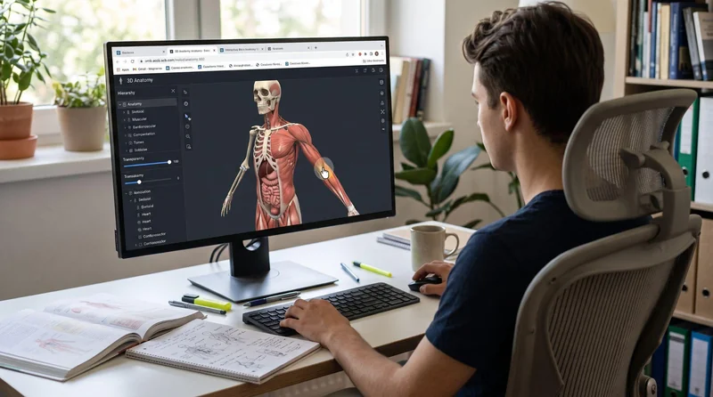
Not long ago, exploring anatomy in detail meant booking time in a dissection lab, sitting at a high-end workstation, or installing expensive software. Today, a similar model can open in a browser tab on a laptop or phone.
This shift changes who gets access to anatomy. Medical students can review a structure on the way to class, clinicians can show patients what is happening inside their bodies, and training teams can share content with a single link. Studios that create 3D medical animation, such as VOKA.io, build detailed anatomical assets that now move from rendered video into interactive web viewers.
Here is what changed, and where browser-based interactive anatomy is already useful.
From Dissection Labs and Workstations to a Browser Tab
Digital anatomy has a longer history than most people think. In November 1994, the U.S. National Library of Medicine released the Visible Human Male, a set of CT, MRI, and photographic cross-sections of a donated body. A female dataset followed in 1995. According to the NLM project page, the male dataset alone was about 15 GBs. In the mid-1990s, working with data of that size required specialist computers and technical staff.
For the next two decades, interactive anatomy stayed close to institutions. Universities bought dedicated software licenses, and some invested in large touchscreen tables for virtual dissection, where students remove layers of a digital body instead of a cadaver. These tools were powerful, but they were tied to a specific room or device.
The browser removes that limit. A web-based 3D anatomy model needs no installation and works across operating systems. A single link can reach every student in a cohort or every patient in a clinic.
What Changed: the Technology in Plain Language
Three changes made browser-based interactive anatomy practical.
- Browsers learned to use the graphics chip. WebGL, introduced in 2011, lets a web page draw 3D graphics with the device’s GPU, the same chip that runs video games. Its successor, WebGPU, now ships in current versions of Chrome, Edge, Firefox, and Safari 26. WebGPU gives developers more efficient access to modern graphics hardware. That helps with larger models, more realistic lighting, and heavier tasks such as displaying 3D volumes built from CT or MRI scans.
- 3D files got smaller and smarter. Formats such as glTF package geometry, materials, and structure names in a compact file designed for fast loading. Compression reduces the download size even further.
- Everyday devices caught up. Mid-range phones and laptops now include graphics hardware that handles real-time 3D, and faster mobile networks make larger downloads less painful.
Together, these changes mean a well-optimized model can load in seconds and rotate smoothly under a fingertip.
Digital Anatomy and Virtual Dissection in Medical Education
Browser-based 3D has the clearest track record in medical education technology. Anatomy is spatial by nature, and flat textbook images make it hard to see how structures relate to one another in depth.
Research supports the move to 3D. A 2015 meta-analysis by Yammine and Violato, covering 2,226 participants, found that 3D visualization tools led to better spatial knowledge and higher factual knowledge scores than other teaching methods. Learners also reported greater satisfaction with the tools.
In practice, browser-based digital anatomy supports learning in several ways:
- Students rotate, hide, and isolate structures to understand depth and relationships.
- Virtual dissection lets them remove layers in order and repeat the process as often as needed.
- Instructors share the same model with a whole class, in the lecture hall or at home.
- Quizzes ask students to find a structure on the model instead of picking a name from a list.
Leggi anche:
Most educators see these tools as a complement to cadaver dissection, not a replacement. Hands-on dissection still teaches tissue texture, anatomical variation, and professional values that a screen cannot fully reproduce.
Clearer Conversations Between Clinicians and Patients
Interactive anatomy also helps outside the classroom. Many patients struggle to picture a diagnosis from words alone. A clinician rotating a 3D model of the spine to show where a disc presses on a nerve can explain the problem in seconds.
Browser delivery makes this easy to repeat. The same model can appear on a tablet during the consultation, on the clinic’s website, and in a follow-up email the patient opens at home with family.
Interactive models and 3D medical animation work well together. An animation shows how a condition develops or how a procedure works, step by step. An interactive model then lets the patient explore the anatomy at their own pace and return with better questions.
Faster, More Flexible Professional Training
Training teams use browser-based anatomy to reach people who cannot gather in one room. Nursing educators can assign a short module before a skills session. Medical device companies can show sales and clinical staff exactly where an implant sits and which structures surround it. Surgical residents can review the anatomy of an approach on a phone the night before a case.
Because content lives on the web, updates reach everyone at once. When a model is corrected, or a new procedure is added, nobody needs to reinstall software.
What Still Requires Attention
Moving anatomy to the browser solves the access problem, but it raises new questions:
- Accuracy. A convincing model can still be wrong. Medical experts should review every structure before publication and after each update.
- Performance. Detailed models need careful optimization, or they will load slowly on older phones and school computers.
- Accessibility. Users who rely on keyboards or screen readers need text alternatives and controls that do not depend on a mouse or touch.
- Context. A model without explanation can confuse beginners. Labels, guided tours, and supporting material turn exploration into learning.
Anatomy for Anyone with a Link
Interactive anatomy has moved from a few well-equipped labs to any device with a modern browser. WebGL made this possible, WebGPU is making it faster, and better-prepared 3D models are making it more useful. For educators, clinicians, and trainers, the question is no longer whether 3D anatomy can run on the web, but how to use it well.
Ti potrebbe interessare:
Segui guruhitech su:
- Google News: bit.ly/gurugooglenews
- Telegram: t.me/guruhitech
- Facebook: facebook.com/guruhitechfb
- Instagram: instagram.com/guruhitech_official/
- X (Twitter): x.com/guruhitech1
- Bluesky: bsky.app/profile/guruhitech.bsky.social
- Rumble: rumble.com/user/guruhitech
- VKontakte: vk.com/guruhitech
- MeWe: mewe.com/i/guruhitech
- Skype: live:.cid.d4cf3836b772da8a
- WhatsApp: bit.ly/whatsappguruhitech
Esprimi il tuo parere!
Ti è stato utile questo articolo? Lascia un commento nell’apposita sezione che trovi più in basso e se ti va, iscriviti alla newsletter.
Per qualsiasi domanda, informazione o assistenza nel mondo della tecnologia, puoi inviare una email all’indirizzo [email protected].
Esprimi il tuo parere!
Che ne pensi di questa notizia? Lascia un commento nell’apposita sezione che trovi più in basso e se ti va, iscriviti alla newsletter.
Per qualsiasi domanda, informazione o assistenza nel mondo della tecnologia, puoi inviare una email all’indirizzo [email protected].
Scopri di più da GuruHiTech
Abbonati per ricevere gli ultimi articoli inviati alla tua e-mail.